How do I choose a good doctor?


We all know that the hunt for a new doctor can be a very long and stressful process. So what is the secret to finding a good doctor in a timely manner? Here are some guidelines to make your decision as pain-free as possible:

  • Credentials – Don’t hesitate to verify a doctor’s medical license, certifications and professional society memberships to ensure he is properly trained.
  • Experience – During a consultation with a doctor ask about his medical education and training that is specific to the procedure you are considering. You can also ask how many procedures of this kind he has performed and how often he performs them.
  • Referrals – Ask your friends and family if they have any doctors that they would like to recommend to you. But keep in mind that doctors specialize in different areas and most are not experts in all areas. So be sure to ask your friend or family member which procedure they had with the doctor they are referring you to.
  • Testimonials – Asking a doctor for testimonials from previous patients will help you understand what qualities others liked about him.

You should also consider scheduling consultations with numerous doctors in order to find the best one for you. Doctors may have different techniques that yield different risks, recovery time and results, and it is worth it to weigh all of your options. It is important that you find a doctor that you are comfortable with and trust.

Doctors are people too

Going to the doctor can often times be daunting. Not only are you nervous about finding out whether or not you are healthy, but you also can be afraid that the doctor will judge you on your health, or more likely unhealthy, habits. However, you need to remember, doctors are people too!

Doctors, just like the rest of us, have their own bad habits and are not here to judge us. You should not be embarrassed when you are discussing your health habits with your doctor.

In fact, they appreciate when you tell the truth about your bad habits instead of keeping them a secret. When a doctor knows your true habits, they will be able to better diagnose you and offer the proper treatment options.

Furthermore, you should not fret over discussing “embarrassing” problems, like bodily functions, with doctors. Anything you say will not surprise the doctor. Most likely your doctor has treated several other patients with the same problem.

Remember your doctor is a regular person just like you and you should tell him the truth so you can get the most out of your visits to the doctor’s office!

Hemorrhoid prevention tips


Hemorrhoids are a very common health problem, in which a lump or mass of tissue in the anus contains enlarged blood vessels. They are caused by an increase in abdominal pressure.

People usually experience hemorrhoids because of:

  • Pregnancy
  • Frequent constipation
  • Obesity
  • Persistent diarrhea
  • Rushing to complete a bowel movement

Additionally, hemorrhoids are likely to develop in individual who do not get enough fiber in their diet or who d not exercise frequently. These poor diet and exercise habits can lead to constipation and straining during bowel movements, which cause or inflame the hemorrhoid.

Reducing your risk of suffering from hemorrhoids can be achieved by making a few simple changes to your diet and lifestyle. The following changes should help soften your stool so that you avoid the straining that leads to hemorrhoids:

  • Increase your fiber intake. You should aim to have about 25 – 30 grams of fiber a day to decrease the risk of constipation.
  • Stay hydrated. Drink at least 6 – 8 glasses of water every day to keep your stool soft.
  • Exercise regularly. Staying active helps prevent constipation and reduces pressure on the veins.
  • Go to the bathroom as soon as you have the urge to have a bowel movement. If you wait to go to the bathroom the stool can become dry and harder to pass.

The importance of knowing your family’s health history

Having a good understanding of your family’s health history is essential in disease prevention. Many of the most common and fatal disease can be hereditary, including heart disease, cancer and diabetes.

Knowing your risks allows you take the right lifesaving steps. Together, you and your doctor will be able to take the right precautions to lower your risk of suffering from an illness that a family member has suffered from. Here are some of the key things you should know about your family health:

  • Medical history of relatives on both sides of your family.
  • The age at which a family member’s health problem began.
  • The races and countries of origins of family members.

Learning about patterns or common illnesses in your family will better prepare you to take early steps to reduce the risk of developing those diseases. Preventative measures can include making simple changes that prompt a healthier lifestyle or receiving appropriate medical screenings early and often.

Wireless Capsule Endoscopy

Endoscopy is a procedure that allows medical professionals to look inside your body. Instruments called endoscopes have cameras attached to long, thin tubes – which are inserted into various orifices of the body to examine nearby organs.

Although traditional methods of endoscopy are used in diagnosing problems in all areas of the body, they are most commonly associated with diagnosing problems of the gastrointestinal (GI) tract. Traditional endoscopy procedures are performed on various components of the GI tract to diagnose conditions like ulcers, Gastro Esophageal Reflux Disease (GERD), polyps, tumors or to find the source of intestinal bleeding. While the traditional methods are beneficial in diagnosing problems of most portions of the GI tract, they are not able to access the small intestine, where some types of serious intestinal conditions, like Crohn’s disease, can begin. Further, some sources of intestinal bleeding are found in the lower intestine and would not be visible through traditional methods of endoscopy.

In 2002, the Food and Drug Administration (FDA) approved a highly effective, minimally invasive method of endoscopy that allows physicians to see the lower portions of the GI tract that traditional methods did not. The new method, known as wireless capsule endoscopy, is a camera that is encapsulated in a pill-like covering, which the patient swallows like they would normal medication. As the capsule moves through the GI tract, the tiny camera takes images of the intestinal walls and organs at the rate of 2-4 images per second for up to eight hours. The images are transferred to a device attached to the patient via a belt worn around the waist. Physicians can then review the images and pinpoint any possible problems. Because the capsule passes through the entire GI tract, it offers physicians a glimpse of all three portions of the small intestine, which traditional methods of endoscopy could not do. After about three days, the pill camera will pass through the body during the natural excrement process.

While the wireless capsule endoscopy is a revolutionary approach to endoscopy that medical professionals at La Peer Health Group offers their patients, it is not suitable for everyone. To discuss which method of endoscopy is right for you, schedule a consultation with one of our health care professionals today.

Facelifts and VECTRA Facial Sculptor

Facelifts are a popular surgical method of facial rejuvenation, with just over 100,000 procedures being performed in the United States annually. During the procedure, incisions are made at various points on the face to take the slack out of lax skin and remove fatty deposits underneath. Some tightening of facial muscles is also done during this procedure.

Even though thousands of people opt to have a facelift done every year, there are likely just as many who have considered it, but never gone through with the idea. The most common reason people decide against having the procedure done is the uncertainty associated with the results. The effects of a facelift are permanent, barring additional procedures, and the thought of having to live with a botched facelift or endure another surgery can be overwhelming. Thanks to improved technology in the field of cosmetic surgery, patients can now see what they will look like after  having a facelift, before the first incision is ever made.

The VECTRA® Facial Sculptor is breaking new ground in cosmetic surgery offices across the country and the cosmetic surgeons of La Peer Health Systems are pleased to announce that they, too, are now offering it to their patients. The tool, created by Canfield Imaging Systems of Fairfield, NJ, the world’s leading provider of medical photography systems – uses six cameras with 3D imaging software to produce a real-time 3D image of each patient. When prospective facelift patients come for an initial consultation, they are easily able to point out problem areas they would like to have addressed, using a 3D model of themselves. Then, based on the patients wishes, our surgeons are able to formulate a plan of corrective measures to take and then produce another 3D model depicting what the patient will look like after the corrective measures are taken. By being able to see the end result before starting, much of the fear and apprehension associated with the procedure can be relieved.

If you have been considering a facelift, but were leery of the results, there is no better time to have one than the present. Our team of qualified cosmetic surgeons can help you achieve the results you always wanted, but have been afraid of pursuing. Schedule a consultation with our cosmetic surgery team today and let the VECTRA® Facial Sculptor introduce you to the new you.

Cosmetic Foot Surgery Los Angeles, Beverly Hills

As society becomes increasingly appearance-conscience, people are paying attention to parts of their bodies they had previously overlooked. While cosmetic surgery was most commonly used to improve and enhance the appearance of the face, the popularity of having elective cosmetic procedures has now spread throughout the body – most recently to the feet. A recent survey conducted by the American Orthopedic Foot & Ankle Society (AOFAS) among its podiatric surgeon members found that just over fifty percent of all their patients had inquired about cosmetic procedures for the feet. So why the sudden interest in cosmetic foot surgery? There are two reasons – to improve the appearance of the foot and to correct deformities of the foot.
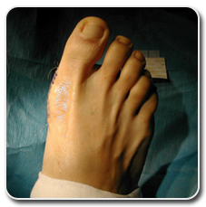
Just like some people think their nose is too big or their breasts are too small, others find faults in their feet. Perhaps they believe their toes are too short, too long, too crooked or that their feet are just too fat.  Cosmetic procedures to improve the appearance of the feet may include removing fat from toes that are too chubby, adding fat to other areas of the feet that may be too thin, surgically narrowing feet that may be too wide or microdermabrasion procedures that smooth the appearance of the feet and remove rough patches of dead or dry skin. Of course, while all of the aforementioned procedures are mere cosmetic enhancements that are purely elective, there are other cosmetic surgeries of the foot that improve that improve the appearance of the foot while correcting an underlying condition that causes the altered appearance.

Conditions like hammertoes, bunions, misaligned or broken toes and other mild deformities of the feet can not only detract from the appearance of the feet, they can also pose mobility problems in the future if left untreated. Walking may become increasingly difficult or even painful if these deformities are not addressed by surgical intervention. The cosmetic benefits of this type of foot surgery are just a perk, not the goal of the procedures.

Most cosmetic surgeries of the feet can be done on an outpatient basis where the patient goes home the same day of the procedure. Depending on what they had done, aftercare can range from hardly any limits at all imposed on activity to the need for crutches for several weeks post-op. As with any surgery, there can be some pain and discomfort after the surgery, which are both easily treated with medications.

If you have been considering cosmetic foot surgery to correct a deformity of the foot or just for cosmetic enhancement only, we invite you to schedule a consultation with our Beverly Hills podiatrist, Dr. Jamshidinia. He can create a plan of treatment, based on your wishes and the findings of the consultation, that will help you put your best foot forward in all that you do.

Email La Peer Health Systems at [email protected] today to get more information about cosmetic foot surgery procedures.

To learn about other cosmetic foot procedures offered at La Peer, please visit BunionDr.com, our site dedicated to bunion treatment and cosmetic foot procedures.

By:

Next, read about Bunion Surgery.

Bunion Surgery Beverly Hills Los Angeles

A bunion is the term given to a knot that appears on the side of the foot, just to the side of the lowest joint of the largest toe. The knot is made up of bone and soft tissue and appears when the largest toe has been pushed inward toward the other toes for an extended period of time. Heredity plays a role in bunion formation, as do illnesses such as arthritis and polio. However, the leading cause of bunions is wearing ill-fitting shoes where the toes are cramped together. Since women are more likely to wear poorly fitting shoes because of heels and such, bunions are more prominent in women. In fact, the American Orthopedic Foot and Ankle Society found that over 85 percent of women in the U.S. wear shoes that are too small and over 50 percent have bunions.

Bunions usually do not become painful unless they are allowed to progress. If they do begin to cause pain, most of the time simply changing the style of the shoe to give the toes more room works wonders in rectifying the problem. However, in the most severe and painful forms, bunions can sometimes require surgical intervention.

Bunion surgery is broken into two main categories: head procedures and base procedures. In head procedures, surgery is done on the joint of the largest toe whereas base procedures are done on the joint behind the largest toe. There are about a hundred different sub procedures that can be formed on various sections of the toe, due to the number of components in each. Not every procedure will work to correct everyone’s problem as no two patients are the same. Aftercare for bunion surgery usually involves the use of crutches for two weeks, keeping the foot dry and elevated during those times and staying off of the affected foot as much as possible in the days following the surgery. Stitches will be removed one week after the procedure, at which time the surgical dressing will also be changed.

There are some caveats that patients should be aware of when it comes to bunion surgery. It should only be performed by a qualified orthopedic surgeon. Patients should be leery of any procedure that promises to provide a quick fix or instant relief to their bunion problem. Due to the intricacy of the structure of the small bones in the toe, bunion surgery is a delicate procedure that should not be taken lightly as not to cause more harm than good. Bunion surgery is meant to correct the deformity and the pain in the toe, not as a cosmetic procedure. So if a surgeon is promising you a “perfect foot” after you have this procedure done, it should serve as a red flag that you may want to look for another surgeon.

If you’re interested in learning more about bunion treatment at La Peer, you can visit our Bunion Surgery Center of Excellence website.

By:

Overactive Bladder

A symptom of an overactive bladder can be the sudden urge to urinate . The urge may be difficult to suppress, which can lead to the involuntary loss of urine, also known as  incontinence. For those who aren’t affected by this condition, an overactive bladder may seem like a minor inconvenience, but for the 25 million Americans that are living with the effects, an overactive bladder can greatly reduce one’s quality of life.

People who are living with an overactive bladder may feel embarrassed, isolate themselves, or limit their work and social lives. They would rather be alone than be caught in a compromising and potentially embarrassing situation due to their bladder problem. This isolation can lead to other, more serious conditions, such as depression.

Overactive bladders are often a side effect of another condition and not a condition within themselves. There are many factors that can lead to an overactive bladder. Some of them the patient has control over while others, they may not. (ameriseed.net) Medical conditions like diabetes, urinary tract infections, bladder and kidney stones, Parkinson’s disease and other neurological disorders can all produce an overactive bladder. Sometimes it happens that once these underlying conditions have been addressed and treated, the overactive bladder problem fades away on its own. There are some behaviors that cause an overactive bladder that the patient must learn to control themselves, such as excessive fluid intake and consumption of too much caffeine or alcohol. Overactive bladders are common in older people, only because they are more susceptible to the underlying conditions that cause them. Overactive bladders are not part of the natural aging process.

There are a number of different methods that a physician will test a bladder for over-activity He may use dyes and imaging devices or various types of bladder stress tests to see how the bladder performs. Once the cause and severity of the overactive bladder has been determined, the doctor will then prescribe a treatment plan that is usually a combination of different methods. Medications combined with bladder re-training exercises are the most common form of treatment for overactive bladders. These re-training exercises may consist of pelvic floor exercises or simply creating a new routine for going to the bathroom in order to put the bladder on a schedule.

Every patient and every bladder is different. This is why a treatment that works for some may not work at all for someone else. If you are experiencing problems due to an overactive bladder,  you don’t have to suffer in awkward silence any longer. Schedule a consultation with our staff to find out which treatment options are available to you so that you can take back control of your life – and your bladder.

Pterygium Excision

A pterygium is a wing-shaped growth that can occur on the eye and eventually cover and alter the shape of the cornea. Pterygia consist of abnormal conjunctival tissue (the membrane that covers the eye) .  Pterygia can be small or grow large enough to affect a person’s vision. It is not clear why pterygia form, but they are twice as likely to occur in males than in females and are most often found in people who live in eye-irritating environments, such as windy climates, smoky places or sandy tropical locations.
While pterygia aren’t dangerous, if large enough, they can obstruct a patient’s vision. The most commonly reported symptoms of pterygia are irritation, redness, dryness, tearing and the sensation of having something stuck in the eye. In most cases, it’s the symptoms of pterygium that are treated, not the pterygium itself. Eye drops and other medications are given to alleviate the dryness and other bothersome effects of the pterygium. Surgical excision of the pterygium is reserved for the most severe cases.

Surgery to remove the pterygium is done on an outpatient basis under local anesthesia. Patients usually go home the same day and are in very little pain. During the recovery period, the patient may wear a patch to protect the eye and apply drops to the eye or ointments in order to speed up the healing process. Redness and irritation may be present as the eye heals from the surgery.

Because the symptoms of pterygium closely resemble the symptoms of other, more serious eye diseases, proper diagnosis by a qualified ophthalmologist is very important. If you are experiencing any of the symptoms associated with pterygium, schedule a complete consultation with one of our qualified ophthalmology staff. Our professionals can determine whether you might be a candidate for pterygium excision and decide which other avenues of treatment may be available to you.

Colorectal Cancer Screening

Colorectal cancer is a cancer that affects two components of the lower digestive system – the colon and the rectum. Colorectal cancer usually presents in the form of per-cancerous polyps in the colon or rectum. Because they are still in their per-cancerous or benign state, these potentially deadly polyps often produce no symptoms until they have become malignant and possibly spread to other organs in the body.

Colorectal cancer is the third most common type of non-skin cancer in both men and women. It is the second leading cause of cancer-related deaths in the United States. Nearly thirty thousand new cases of colorectal cancer are diagnosed in this country every year. Colorectal cancer affects men more than it does women and is most common in people over the age of fifty. People with a family history of colorectal cancer, other hereditary diseases or colon conditions such as Crohn colitis are more likely to develop colorectal cancer than those who do not.

Because of its often silent nature, regular screenings for colorectal cancer are imperative for early detection, when the cancer is in its most treatable stages. Doctors recommend that people with no family history of colorectal cancer or hereditary conditions that can lead to it be screened at age fifty and again at regular intervals as directed by their physicians. People who do have a family history of colorectal cancer or that have any type of inflammatory bowl disease are encouraged to begin their testing earlier in life and to have the screenings done more frequently.

Colorectal cancer screenings are usually done by inserting an imaging device into the rectum that will allow doctors to examine the rectum and the lower part of the digestive tract. There are a few different imaging devices and tests that can be utilized, with each one allowing access to different parts of the lower digestive system. If a problem area is detected, the doctor will order additional testing and perhaps a biopsy of any suspicious growths. Depending on the results of the biopsy, the doctor will recommend a course of treatment if the growth is found to be cancerous. The treatment will depend upon the location of the cancer, its stage and the patient’s overall health, among other factors.

Even the most knowledgeable doctors using the latest imaging equipment are capable of overlooking suspicious growths. This is why regular screenings for colorectal cancer are so important. Early detection can literally mean the difference between life and death. If you are aged 50 or older or have some of the preexisting conditions as described above and have not been screened for colorectal cancer recently, we urge you to contact our staff to make an appointment to be screened as soon as possible.

Hammertoes, StayFuse Intramedullary Fusion System

Hammertoe is a condition where one or both muscles of the toes is severely bent, causing the toe to take on a deformed appearance that resembles a ’hammer’. Hammertoes generally start out as mild defects, which are often aggravated by the pressure of wearing shoes. Over time, the joints can become more and more bent, resulting in a more extreme – and sometimes debilitating – deformity. Left untreated, hammertoes will become rigid over time and will require surgical intervention to correct.
There are a variety of causes of  hammertoes, but the most common is an imbalance in the muscles or tendons. Toes that are too long for shoes or injury and trauma to the toe are other reasons this condition may develop. There are a number of non-surgical options that can treat a hammertoe in its early stages. Some of these are injection therapy, wearing corrective footwear and applying splints to realign the affected toe or toes.

If these methods do not alleviate the condition, surgical intervention may be a patient’s last resort. One of the surgical methods that podiatry specialists at the La Peer Health Systems use is the StayFuse Intramedullary Fusion System by Tournier. The StayFuse™ device consists of an interlocking two piece titanium screw that snaps together for the fusion of small bones and replaces the 1.1mm K-wires used in traditional methods.

To correct a hammertoe using the StayFuse method, the first and second bones of the toe are fused together at the point where the tendon was overstretched and caused the crooked appearance. The devices are color-coded by size, decreasing length and circumference for each toe to accommodate the change in bone size. The benefits of the StayFuse Intramedullary Fusion System over other methods of surgical repair of hammertoes is a decreased chance of infection, no post-op implant exposure and no disruption of healthy joints.

If you’ve been suffering from the pain that’s often associated with hammertoes, the good news is you don’t have to suffer any longer. The specialists at La Peer Health Systems can help you decide which treatment method may be right for you by evaluating your condition and concerns. Contact our office to schedule a consultation with a member of our podiatry team today.