Postoperative Care for Minimally Invasive Spine Surgery

The management of the needs and activities following a minimally invasive spine surgery evolve with time. Patients will have some expectations during this stage. The spine surgeon will have various recommendations based on his or her preference, the need of the patient and the type of minimally invasive spine surgery. Immediately after the surgery, pain medication is administered. This is usually given by injection or in the vein. Within two days, as the pain lessens, this can be modified to oral medication.

Recovery in preoperative symptoms is attained in 80% of patients. Back pain and function improvement continues up to a couple of years following the minimally invasive spine surgery. The basis for the improvement would be:

  1. nerve compression recovery
  2. continuing fusion mass maturation
  3. muscle conditioning after the surgery

Recovery after a minimally invasive spine surgery can be affected by smoking. Nicotine is like a bone poison that inhibits bone fusion. According to a study on bone mass fusion with spinal fusion patients depicts that there is lower nonunion rate to patients who do not smoke than those patients who smoke. The nonunion rate for those who quit smoking is slightly higher.

Occupational therapy, exercise and physical therapy may be needed to aid patients in regaining strength and function. This could start in the hospital or be suggested at a later time

Like other forms of back pain treatment, the more effort exerted by the patient, the better the whole result of minimally invasive spine surgery.

Non Surgical Treatment Options for Gastrointestinal Problems

There are a number of problems that usually affect the gastrointestinal tract. A few of them are often short lived; however, they cause discomfort and usually distressing. A lot of problems that affect the gastrointestinal tract can be resolved by improving the lifestyle and following doctor’s prescriptions.

Diarrhea- This disorder is treated while ensuring that dehydration will not happen again and decrease the rate of food movement through the bowel.

  1. Kaolin- This is a known remedy due to its reputation. This medication is available in tablet or liquid form. Anything in the bowel that may cause diarrhea is absorbed by the kaolin.
  2. Oral rehydration sachets- This must be the main treatment for diarrhea. Only these sachets replenish the salts in the body that it lost along with water during the disorder.
  3. Loperamide- This functions like Morphine but it is more effective. An episode of diarrhea can usually be treated with a couple of capsules as one dose.

Constipation- The main goal of treating constipation is to make some improvements in the bowel movement. This is made possible by taking in laxatives. Laxatives come in different types.

  1. Osmotic Laxatives- These functions by pulling water into the bowel which will soften the bowel content and minimize constipation.
  2. Stimulant Laxatives- These medicines are helpful in leading the passing of a movement and relieve constipation. They will irritate the bowel lining and cause bowel muscle contractions.
  3. Lubricating Agents- These agents line the bowel with lubricants to ease the passing of a movement.

Indigestion-Treating this disorder will vary according to its symptoms. Its treatment is aimed at removing the excess acid. Here are several ways to do this.

  1. Alginates- They react with acid and come up with a rafting effect. They are available in liquid or tablet form.
  2. Antacids- They contain ingredients that neutralize stomach acids

Gastrointestinal Problems: Surgery

Hand-assisted laparoscopic surgery (HALS) and laparoscopic surgery are minimally invasive procedures that are usually used to diagnose disorders in the gastrointestinal tract.
Some of the conditions that are treated at La Peer Surgery Center are as follows. Please contact us if you think you might be suffering from any of the signs and symptoms listed above.

Medical Conditions
Cirrhosis of the Liver
Inflammatory Bowel Disease
Fecal Incontinence,
Colorectal Cancer
Peptic Ulcer Disease
Ulcerative Colitis
Irritable Bowel Syndrome
Chronic Viral Hepatitis
Constipation treatment
Celiac Diet
Lactose Intolerance
Anal Warts
Pancreatitis treatment
Inflammatory Bowel Diseases
Hemorrhoid treatment
Severe Constipation
GERD

Orthopedic Surgery


INTRODUCTION
To correct their musculoskeletal deformities such as fractures and fragile bones, patients who experience these kinds of conditions resort for an Orthopedic Surgery. It is a surgical treatment performed by a medical specialist specifically an orthopedic surgeon, who assesses and cures problems involving the bones, joints and the ligaments. Common surgical procedures include ORIF (Open Reduction with Internal Fixation), Closed Reduction with Internal Fixation and Arthroscopic surgeries of the knee. It may also treat problems involving the nervous system such as Muscular Dystrophy and Cerebral Palsy. It also remedies different skeletal deformities in the spinal cord, such as following an injury. These problems may be congenital in nature, through an injury or declining function associated with age. It could be classified as acute, where an injury or trauma causes the alteration of function, or chronic such as age-related changes.

In the past, orthopedic surgeons treated spine and leg irregularities in children where they apply braces to strengthen the bones of the child. It was where the name Orthopedics originated, from two Greek words “ortho” meaning straight, and “pais“, the Greek name for child. Through the advents of anesthesia and the application of aseptic techniques, the work of orthopedic surgeons gradually shifted to associated nerves and tissues of different bones.

Although several orthopedic surgeons undergo specialization in particular surgeries such as spinal disorders and in hand and joint replacement surgeries, most of them are still practicing general surgery. Others might specialize in treating injuries in trauma units and emergency rooms, a field called as trauma medicine. Some of them might even go beyond their field and collaborate with podiatrists, geriatric specialists and other related medical allied groups. An emerging field of orthopedics is Sports Medicine, which are composed of board certified orthopedic surgeons.

Description
A variety of extensive procedures are offered by orthopedic surgeons, such as amputations, traction, spinal fusion, hand reconstruction and joint replacement. Strains, sprains, dislocations and broken bones are also treated by orthopedists. Specific procedures are done by orthopedic surgeons such as arthroplasty, arthroscopic surgery, fracture repair, traction, fasciotomy, and bone grafting, to name some of them. Different hospitals provide work for these orthopedists and incorporate them as part of the surgical team along with the surgical nurse and anesthesiologist. Before the orthopedic surgery is done, a choice of general, regional or local anesthesia could be used. A surgical team may compose of a solo practitioner or a group of two to six orthopedists.

The orthopedic surgeons’ job includes adding screws, wires, nails, pins, prosthetics and tongs inside the body. In this way, proper alignment of the skeleton is achieved and damaged bone or tissue is substituted. Enormous strides have been made with the introduction of recent devices such as artificial limb and joints with the availability of new materials used to restore damaged bones and connective tissue. Advancements in plastics and metal technology enable orthopedists to mimic the natural functions of bones, joints and ligaments and achieving greater accuracy in restoring damaged parts to their original range of motion.

Orthopedic surgeons make use of physical, medical and rehabilitative methods. In order for them to remove dead cartilage and other debris in the affected area, they often resort to laser surgery. If the fracture or dislocation is sports-related, these surgeons would elect to procedures such as shoulder arthroplasty when the fractured part is on the shoulder, or replacing the ball and socket joint for osteoarthritis.

Diagnosis and Preparation

Other doctors such as emergency doctors and primary care physicians may refer their patients to an orthopedic specialist, preferably a surgeon. They work alongside other healthcare teams that include family physicians and rheumatologists. Before the orthopedic surgery is done, candidates for the operation would have to a screening procedure to determine the appropriate procedures to be done. The procedure to be done greatly lies on the patient’s age, underlying orthopedic condition, physical health in general and the impact of joint disability to the patient’s daily activities of living. To ensure greatest function, the timing of the procedure must be crucial. Surgery should be performed before surrounding muscles become contracted and atrophied and the occurrence of serious structural abnormalities. The orthopedist makes a thorough evaluation of the client to ensure that the correct procedure is to be performed.

A series of diagnostic tests include X-ray, Computed Tomography Scans and Magnetic Resonance Imaging (MRI), along with series of blood tests for donor compatibility, diagnostic arthroplasty and myelograms. The history of the deformity together with the patient’s history of treatments is determined by the orthopedist. Allowing the affected area to rest prior to surgery may be carried out. Patients who will go through orthopedic surgery must also pass standard serum and urine tests prior to the operation, and may undergo electrocardiogram (ECG) and other necessary tests before the surgery commences. Patients scheduled for an operation may donate their own blood to make it as a reserve for major surgeries, since blood loss is common. It also minimizes the risk of blood transfusion reactions.

Minimally Invasive Spine Surgery: Description

Minimally invasive spine surgery is an endoscopic operation that utilizes specialized instruments that are entered into the abdomen or chest through tiny incisions in order to reach the spine and do the required surgery. This procedure involves the use of a specialized video camera as well.

For decades, endoscopic methods have been utilized but to diagnosis certain conditions only. In the later part of the 1970 and early part of the 1980s, there were advancements made to endoscopic techniques which allowed a diagnosis to be made and the disorder could be remedied. These techniques that are adopted in other surgical requirements have been developed to treat spine disorders.

However, a minimally invasive spine surgery is not perfect for everyone. A surgeon must completely assess each patient to identify their qualification for the surgery. In some cases of scoliosis, degenerative discs, kyphosis, herniated discs, fractures and infections, minimally invasive surgeries may accelerate recovery, alleviate post-operative pain and enhance the final result. The different types of minimally invasive spine surgeries are:

Spinal Tumor Decompression– This is performed to remove the whole tumor or part of it from the spine. This procedure decompresses or relieves spinal cord and nerve root pressure, thereby alleviating pain and other potential symptoms.

Minimally Invasive Lumbar Spinal Fusion-This procedure is aimed at fusing the two vertebrae to stop any motion between them. Removal of the intervertebral disc or bone spurs may minimize nerve pressure. When the two vertebrae are fused, the bone spur formation will be stopped at that certain location, further alleviating possible nerve injury and pain.

Discectomy is a procedure that is a removal of a herniated disc material that presses on a nerve root or the spinal cord. Before the removal of the disc material, a tiny piece of vertebral bone is removed. This is to enable the surgeon to have a better view of the herniated disc.

Benefits of Minimally Invasive Surgery
Compared to the conventional spine surgery that needs long incision and recovery period, minimally invasive surgery utilizes an endoscope that is inserted into a tiny incision. The endoscope is attached to a small video camera that projects the inner view of the body onto the operating room’s television screens. After the procedure, sutures are used to close the tiny incisions which will be covered with surgical tape. They will be almost invisible after a few months.

The fact that minimally invasive spine surgery holds a minimized risk of complications and alleviates the need for a longer stay in the hospital indicates that there could be cases that such surgical procedure can be performed on an outpatient basis. Obviously, this demonstrates substantial advantage for both patient and doctor in terms of cost effectiveness and safety. These procedures have become popular due to these mentioned advantages.

Minimally invasive spine surgery techniques use cutting-edge technology to make corrections to chronic spinal conditions without triggering undue injury to the soft tissues that surround it. Because of these highly specialized instrumentation and tools, guided by enhanced features like nerve monitoring and computerized navigation, spine surgeons are able to perform the surgery safely and effectively.

Compared to posterior lumbar fusion (PLF), a minimally invasive spine surgery like transforaminal lumbar interbody fusion (TLIF) provides the same postoperative fusion rates and alleviates the quantity of spinal muscle iatrogenic injury. The benefits of minimally invasive spine surgery include:

  1. Lower infection risk
  2. Minimized post-operative  pain
  3. Alleviated blood loss
  4. Minimized scars
  5. Less body trauma
  6. Shorter recovery period
  7. Reduced or eliminated general anesthesia time
  8. Enhanced post-surgical mobility and function

Risks and Complications of Minimally Invasive Spine Surgery

Like any surgical operations, minimally invasive spine surgery has both procedure-specific and general risks. There are also cases that the original planned surgery will not be completed and must have a round of operation or a diversion from the minimally invasive technique to a full open approach.
General Risks:

  1. Adverse reactions to anesthetic
  2. Leg blood clot that can travel to the lungs causing pulmonary embolus
  3. Post-operative pneumonia
  4. Surgical site infection
  5. Blood loss that will require a transfusion

Procedure-specific risks:

  1. Spinal cord or nerve injury that results in pain and paralysis in severe cases
  2. Possible breakage and malfunction of the instrumentation used that may irritate the surrounding tissues
  3. Surgical pain

Surgeons who are performing minimally invasive surgery usually would say that there is a limited opportunity to learn this technique. Post-operative and intra-operative problems are higher when the surgeon is obtaining experience. The technical challenges may be overcome with practice and training but it needs time.

Minimally invasive spine surgery requires advanced techniques, demands specialized equipment, offers decreased visualization and is linked to a limited learning curve.  Compared to conventional surgical methods, this type of procedure is also related to minimized fusion rates and insufficient decompression.

The procedure does not need the utilization of fluoroscopy. This means a low chance of exposure to fluoroscopy. Actually, the risk is more for the part of the surgeon and staff in the operating room who are exposed to every patient. In minimally, invasive spine surgery, there is usually a risk of bone fracture, nerve damage, screw malfunction and blood clot formation.

Hernia: Treatment

Hernia: Treatment

Hernia treatment can be surgical or non-surgical based on its severity. Usually, non-surgical treatments help ease the discomfort and pain that resulted from a hernia while surgical treatments are used for extreme and severe cases of hernia.

Non-Surgical Hernia Treatment

  1. Henria Belts– These are lightweight belts that are made to give easiness movement during recovery. They are designed to be worn over belts with straps that can be adjusted and are lined with foam for additional comfort.
  2. Trusses- These come as umbilical and spring trusses. The umbilical truss is worn around the midsection while the spring truss is used around waist to act as a support.
  3. Hernia Briefs– These types of briefs look just like any ordinary briefs except that they are made with foam pads and spandex to give additional support.
  4. Bindings- Bindings for the abdomen are elastic and offer constant compression and abdominal muscles support.

Surgical Hernia Treatments
The surgical operation for hernia involves the replacement of hernia contents in the abdominal cavity and repair of the defect. Usually, a prosthetic material must be fitted to allow the safe repair. The operation needs anesthesia administration and it is likely that after or during the operation, blood or blood products might be used.
If you would like to come in to La Peer for a consultation, please contact us via the contact form or give us a ring.

Hernia: Signs and Symptoms

Hernia symptoms vary according to the structure and cause involved. A lot of hernia cases start as small and unnoticeable breakthroughs. It may occur as soft lumps found under the skin at first. Usually, this stage does not involve any pain. However, there will be gradual increase of the pressure in the internal content against the weak wall and the lump size increases. Other symptoms of hernia include:

Pain- Hernia comes with pain due to some reasons that include stretch of the tissue around and at the hole. This is also likely to occur due to damage and irritation to nerves in the area as a result of the pinch and push of hernia nerves.

Bulge- This becomes evident when abdominal cavity contents are pushed through the hole or the hernia defect. This location of the bulge will vary based on the location of the defect or hole and the hernia type.

Localize pain– This kind of pain is often the result of tearing and stretching of the abdominal wall tissues like the tendon and the area muscle. When the bulge increases, the pain is also expected to get more intense.

Generalized pain– Due to the incarceration or entrapment of hernia contents, the blood supply of the intestine is likely to shut off or be compromised. The tissue contained in a hernia will then be strangulated causing the tissue to die and cause additional pain that will become generalized pain throughout the abdomen. The pain is expected to be unremitting and severe.

Hernia Description
Hernia causes discomfort and pain as well as lessens general mobility. It is not a type of disorder that does not heal by itself despite curability of other forms using external manipulation. A hernia is likely to cause complications that are risky depending on the structural solidity through which it is protruding and the nature of the protruding organ.

There are different types of hernia’s as follows:
Abdominal wall hernia– This type of hernia is also called a ventral or epigastric hernia. This is not a very common type of hernia. This type also includes umbilical and inguinal hernias.

Direct inguinal hernia– This type of hernia can affect both men and women. The intestinal loop makes a swelling in the inside part of the groin’s fold.

Indirect inguinal hernia– This type is possible in men only. An intestinal loop passes through the canal from where a testis goes down the scrotum during childhood years. If this hernia is ignored, it may increase in size progressively and cause the scrotum to expand.

Umbilical hernia– Both men and women can be affected by this type of hernia. A loop in the intestine protrudes through a weak abdominal wall at the navel.

Hernia: Causes
Although some abdominal hernias may occur since birth, other types of hernias are developed at a later stage in life. There are those that include pathways created during fetal development, existing abdominal cavity opening and abdominal-wall weakness areas. A hernia can form and become worse by any condition that may increase abdominal cavity pressure. Some of these conditions include:

  1. Heavy lifting
  2. Chronic lung disease
  3. Obesity
  4. Straining during urination or bowel movement
  5. Abdominal cavity fluid

Hernia: Diagnosis

It’s likely that hernias are painful and definitely affect the quality of life. Because of this, it is necessary to visit a physician immediately once you can feel the potential presence of a hernia in the groin and abdominal area. The doctor will use various ways to identify if there is a need for hernia diagnosis so they can continue with an efficient and effective treatment course.

In various cases, hernia diagnosis is needed based on simple physical examination. While considering the type and size of protrusion, the physician may diagnose the condition by pressing gently on the affected area.  However, when a hernia is not simple enough to be identified by a physical examination, the physician may use other methods to identify the order of hernia diagnosis. These methods may include ordering a computerized tomography (CT) scan and magnetic resonance imaging (MRI).

Together with these tests, a diagnostic laparoscopy can also be considered by the physician before finally settling on a diagnosis. This test is a minimally invasive procedure that uses general anesthesia. The abdominal cavity will be directly placed with a scope enabling the physician to perform an examination of the abdominal walls. Typically, this method is only used when there are no conclusive results provided by other procedures of assessing the potential presence of a hernia. Moreover, ultrasound can be utilized to detect a femoral hernia and abdominal X-rays can be ordered by the physician to identify the presence of a bowel obstruction.

When the physician has diagnosed a hernia presence, it is likely to start administering the right treatment for the disorder which will vary according to age, size and location of the hernia and the level of discomfort.

Symptoms of Hammer Toes

Podiatry ExamFeet are incredibly important, in that they help you get from place to place. Additionally, in a warm climate like Los Angeles, feet are frequently on display in sandals or at the beach. If you’re suffering from hammertoes, you may experience not only pain or imbalance when walking and performing other activities but also displeasure over your foot’s appearance. This is where the board-certified podiatrists at La Peer Health Systems can help.

Feet are incredibly important, in that they help you get from place to place. Additionally, in a warm climate like Los Angeles, feet are frequently on display in sandals or at the beach. If you’re suffering from hammertoes, you may experience not only pain or imbalance when walking and performing other activities but also displeasure over your foot’s appearance. This is where the board-certified podiatrists at La Peer Health Systems can help.

Hammertoe Anatomy

Hammertoe is a deformity of the foot in which an enlarged middle toe joint causes the toe to bend in a downward direction. It is called hammertoe because the bent toe can resemble a hammer in its shape. Other people will observe that their toe has a claw-like appearance. As the condition worsens, the joint can enlarge further, stiffening as it comes into contact with the shoe.

Causes of Hammertoe

Many cases of hammertoe are caused by improper footwear. Shoes that are too narrow or too high in the heel may cause the toes to bend. Over time, the toes can become fixed in these unnatural positions, and the foot muscles are unable to bend them back into place. Hammertoe can also result from muscle imbalances and other factors, so it’s important to see a skilled podiatrist about your condition.

Hammertoe Diagnosis

Hammertoe is a simple condition for an experienced podiatrist to diagnose. The doctor will examine your foot, looking for toes bent into a claw-like position. Hammertoe may affect any toe on the foot, but it usually impacts the second toe. The doctor may also order x-rays, as hammertoe can involve bone deformity.

Hammertoe Treatment

Hammertoe can be treated with a simple outpatient surgical procedure. The surgeon will relax the tissue of the foot and help reposition the toes to a more normal setting. This relatively easy procedure can relieve the pain and unsightly appearance associated with hammertoe, while leaving the patient free to return to normal activity the very same day. Learn more about hammertoe surgery on our Foot and Ankle Surgery Center of Excellence website.

Hammertoe Surgery Recovery

Hammertoe surgery is an outpatient procedure with a remarkably fast recovery rate. Generally, patients can walk again on the same day they have surgery with the aid of a protective boot. Pain from hammertoe surgery is usually minimal, lasting only 48-72 hours, and most patients tolerate it well with the help of over-the-counter pain medications.

Contact a Surgeon at La Peer Health Systems Today!

From walking to dancing, we use our feet in a multitude of ways, and for this reason proper foot health is essential to living a full and meaningful life. If you think you may be suffering from hammertoe, it is crucial that you contact a skilled podiatrist.

At La Peer Health System’s Podiatry Department, we employ a highly experienced team of doctors and nurses to help with all your foot health needs and are widely regarded as one of the best foot surgery centers in the country.

Hammer Toes: Treatment

Treatment

{loadposition contactformraw}

In children, mild hammer toe can be treated by manipulating and applying a splint in the affected toe. Changes in footwear such as the following tips may help relieve the symptoms:

  1. Wearing shoes that best fit the toe or opting to choose for shoes with wide toe boxes for comfort, and to avoid further progression of the hammer toe.
  2. Avoiding high heels as much as possible.
  3. Wearing insoles might help relieve pressure in the toe.
  4. Using corn pads or felt pads to protect sticking out joints.

A foot doctor (podiatrist) can make foot devices such as straighteners and hammer toe regulators as part of the treatment regimen. These could be bought as well.
Having an exercise regimen could help. Gentle stretching exercises can be done if the toe is not in a fixed position. Picking up a towel with the use of your toes can aid in stretching and straightening the foot’s smaller muscles.

For a severe hammer toe, an operation called osteotomy might be done to straighten back the joint.

  1. The surgery to be done might involve cutting and moving tendons and ligaments.
  2. Sometimes the bones on each joint are needed to be fused or connected to each other.

In most cases, this is an ambulatory surgery and the patient is allowed to go home on the day of the surgery. right after it. A feeling of stiffness may be present in the toe, yet it may go away in a shorter amount of time. If the underlying condition is treated early, surgery is oftentimes unnecessary. Necessary treatments are done to reduce pain threshold and eliminate walking discomforts. If hammer toe is suspected, an appointment with the physician regarding the best treatment options for pain and walking discomforts could also be done. For prevention purposes, shoes that are too short and narrow must not be worn. For children, regularly inspect foot growth if it coincides well with the current shoe size of the child.
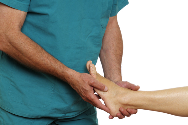
The toes could still be moved at first. But over time, the person might find it hard to move his bigger toes, especially his middle toe.

Presence of Corn
A corn appears in the affected toe. It is caused by rapid cell growth in the toes that is caused by an occurrence where a healing blister leads the formation of a corn as the produced scar tissue is thicker than the normal skin. On the top, a solid corn may emerge and a distal corn at the hammer shaped side.

Callus Formation
A callus, or a thickened portion of the skin, appears on the sole part of the foot.

Pain
With the presence of corn and callus, when tight shoes are worn, it presents a painful experience, more so when walking around. Wearing poorly fitted footwear such as narrow, pointed toe shoes might predispose one to the symptoms, with pain as one of the late signs.

Nail Changes
The nail might split into two or may converge inwards.

Hammer Toes: Description
A Hammer Toe is marked by the contracture of the tendons, ligament laxity and angulations of the second and third phalanges of the toe. It composes of flexion deformities in the proximal interphalangal (PIP) joint of the toe, with a hyper extended metatarsophalangeal (MTP) and distal interphalangeal (DIP) joints. As one of the most painful toe disorders, hammertoe is traced from wearing pointed, narrow toe footwear. Women are more prone to hammertoes than men. Female shoes for most of the time have pointed front portions, and not much wide enough, thus making the foot look round. Hammertoe is created from a blend of factors such as narrow shoes and wider foot or pointed footwear and a rounded foot.

Compression of the feet and constriction of the toes depends on the shoes being used. These may eventually lead to muscle wasting, and decreased motions in the toes. And the result is the toes may have little room to operate. In an ill-fitting shoe, the toe seeks room anywhere it can be found. The pressure is increased, thus leading to the hammer-like shape of the toe.

Due to continuous PIP joint flexion deformity, the occurrence of MTP and DIP compensatory hyperextension might occur. The hyperextension of the MTP joint and the flexion of the PIP joint make the PIP joint move dorsally. This prominence is rubbed against the patient’s shoe, thus the pain is felt. The deformity is flexible and could be passively corrected early but eventually could be corrected as time goes by. Progressive deformities may eventually lead to dislocated joints.

Causes
Most causes of hammer toes come from the person’s selection of shoes, yet other factors also play a role in its formation.

Footgear
The choice of foot gears, not only just shoes but also ill-fitting stockings, tapered toe shoes, pointed toe shoes, tight leotards and snugly pantyhose could all lead to a painful hammertoe. There could even be a possibility of getting hammertoes foot in both feet, since basically these things are worn in both extremities. It might also lead to nerve and joint damage.

Genetic Factors
Some people might be born with a hereditary contracture, but the only thing that increases the risk of familial tendencies is wearing ill-fitting shoes, even though an individual is predisposed to such as a systemic disease like arthritis.

Diagnosis
Usually, a physical examination is done to confirm the presence of hammer toes. The health care provider, such as a physician might find decreased and painful movement of the toes. The patient is asked with the following questions as part of the initial assessment:

  1. A presence of fever or localized erythema. An erythema might suggest phlebitis, gout, osteomyelitis, cellulitis, ingrown toenail and paronychia. Fever may present signs of infection such as osteomyelitis and cellulitis.  
  2. If there is an associative foot deformity. Aside from hammertoe, hallux vagus, hallux rigidus, arthritis and displaced fractures are other foot deformities also assessed.
  3. The presence of palpable peripheral pulses. Diminishing arterial pulses would be a conclusion of arterial embolism, peripheral arteriosclerosis and diabetes.
  4. An associated neurological finding. The presence or loss of sensation of touch and pain should make one a possibility for peripheral neuropathy or carpal tunnel syndrome. Morton’s Neuroma is also associated with numbness or loss of sensation in the 3rd and 4th toes.

Diagnostic Tests
A series of routine tests are done, including a CBC, sedimentation rate, chemistry panel, VDRL tests and X-Rays of the foot. If there are diminishing peripheral pulses, Doppler tests and angiography are measured. Venography is done if there is a presence of diffuse swelling and erythema. Bone and CT scans as well as arthroscopy are considered if results are found out to be negative. To diagnose stress fractures, a MRI may be needed. With the use of quantitative scintigraphs, abnormal weight distribution in the toes may be diagnosed.

Treatment

{loadposition contactformraw}

In children, mild hammer toe can be treated by manipulating and applying a splint in the affected toe. Changes in footwear such as the following tips may help relieve the symptoms:

  1. Wearing shoes that best fit the toe or opting to choose for shoes with wide toe boxes for comfort, and to avoid further progression of the hammer toe.
  2. Avoiding high heels as much as possible.
  3. Wearing insoles might help relieve pressure in the toe.
  4. Using corn pads or felt pads to protect sticking out joints.

A foot doctor (podiatrist) can make foot devices such as straighteners and hammer toe regulators as part of the treatment regimen. These could be bought as well.
Having an exercise regimen could help. Gentle stretching exercises can be done if t
he toe is not in a fixed position. Picking up a towel with the use of your toes can aid in stretching and straightening the foot’s smaller muscles.
For a severe hammer toe, an operation called osteotomy might be done to straighten back the joint.

  1. The surgery to be done might involve cutting and moving tendons and ligaments.
  2. Sometimes the bones on each joint are needed to be fused or connected to each other.

In most cases, this is an ambulatory surgery and the patient is allowed to go home on the day of the surgery. right after it. A feeling of stiffness may be present in the toe, yet it may go away in a shorter amount of time. If the underlying condition is treated early, surgery is oftentimes unnecessary. Necessary treatments are done to reduce pain threshold and eliminate walking discomforts. If hammer toe is suspected, an appointment with the physician regarding the best treatment options for pain and walking discomforts could also be done. For prevention purposes, shoes that are too short and narrow must not be worn. For children, regularly inspect foot growth if it coincides well with the current shoe size of the child.

Symptoms of Gastrointestinal Problems

Gastrointestinal problems will have disorders in particular organs that include intestines, stomach, gallbladder, pancreas, liver and the leading ducts between organs. Gastrointestinal problems may cause an extensive array of symptoms and assessment of a physician is important to identify the causes of the problems.

Heartburn
Those who are experiencing heartburn that stays after modifications to dietary habits and home treatment might be having a gastrointestinal disorder. People with Gastroesophageal reflux disease (GERD) will experience constant heartburn, bad taste and indigestion. People who have gastric disorders like ulcer may have heartburn too that may come with indigestion.

Bowel Changes
People who are experiencing gastrointestinal problems can feel changes in their bowel movements. They might be having constipation and diarrhea from gastrointestinal problems like diverticulitis and hepatitis. They are also likely to experience bloody stools, light colored stools and mucus-filled tools.

Gas
People who have gastrointestinal problems may experience uncontrollable or frequent gas. Peptic ulcers, irritable bowel syndrome and celiac disease may cause excessive and painful flatulence. These people with such problems may have constant belching that may happen frequently after taking in some fibrous foods or fermented or carbonated beverages.

Pain
Those with gastrointestinal problems like diverticulitis are likely to have abdominal and stomach tenderness. Other gastrointestinal problems including Crohn’s disease can cause cramping and pain during a bowel movement. People with peptic ulcers can experience frequent stomach discomfort and pain that becomes worse when they lay down.

Common Gastrointestinal Problems
There are millions of people in the world who are experiencing gastrointestinal problems that range from heartburn, indigestion, ulcer, diarrhea, gas, irritable bowel syndrome (IBS). These problems can cause discomfort and may be frustrating.

These problems affect both men and women of all ages, though they are more common in women. Acute problems can be treated easily and changes to lifestyles are usually recommended for these.

Constipation– This is described as a decrease in the volume and frequency of bowel movements. This usually comes with discomfort that passes a motion or permanent lower abdomen discomfort.

Indigestion– This problem is associated with too much stomach acid which could be a result of a huge alcohol intake or greasy and spicy meal.

Diarrhea – An increase in frequency of bowel movements and the discharge of loose watery stools. A person  with diarrhea may also have abdominal cramps and nausea.

Irritable bowel syndrome (IBS) – This is a functional bowel disorder which means that the function of the digestive tract is impaired. A person may experience abdominal bloating, pain and gas, mucus in stool. irregular bowel habits with constipation, diarrhea or both.

Ulcer– This condition is described by a small hole or erosion in the gastrointestinal tract.

Gas– The presence of gas in the digestive tract is demonstrated by abdominal pain, bloating and bleaching.

Causes of Gastrointestinal Problems
Complaints associated to digestive system usually occur in an ambulatory clinic setting. The uneasiness or discomfort can be stress or diet or psychogenic related. It is possible for such systematic disease to manifest as gastrointestinal symptoms. The majority of problems tend to overlap with organic and functional causes. The following are the major causes of gastrointestinal problems.

  1. Functional Disturbances
    1. Deficiency of secretory IgA
    2. Deficiency of stomach acid
    3. Insufficiency of pancreatic digestive enzymes
  2. Microbial imbalances
    1. Viral infections
    2. Parasitic infections
    3. Bacterial overgrowth
    4. Normal flora imbalances
  3. Destructive agents
    1. Excessive alcohol
    2. Environmental chemicals
    3. Food vulnerabilities
    4. Pharmaceutical agents
    5. Oxidative stress

Diagnosis of Gastrointestinal Problems

Diagnosing gastrointestinal problems will vary according to the type of gastrointestinal disorder. Usually, the symptoms, medical history and physical examination of patients will be the basis of the diagnosis.

Constipation- The doctor may perform an examination to the thyroid gland and the nervous system as well as check medications to determine that such constipation is not a side effect of something else. Typically constipation cases are either diagnosed in a medical clinic or self-diagnosed.

Inflammatory Bowel Disease (IBS) – It is not easy to diagnose inflammatory bowel disease and it is usually mistaken for uncomfortable bowel syndrome. An irritable bowel syndrome does not demonstrate the inflammation that is typical to ulcerative colitis and Crohn’s diseases. The diagnosis for IBS is performed according to the symptoms, physical examination and medical history of the person.

Diarrhea- Apart from asking about the medical history, performing physical examination and testing routine urine, stool and blood tests, the physician may recommend colonoscopy and sigmoidoscopy test for a case of diarrhea.

Indigestion- The physician is likely to ask about the symptoms of indigestion, any taken medication and the lifestyle of the patient. The GP may further perform other examinations like pressing gently some areas of the body to know whether or not it is painful.

Hemorrhoids- This disorder is diagnosed by performing a rectal examination. For internal hemorrhoids, a physician will insert an anoscope. The entire rectum will be examined by using a proctoscope. Sigmoidoscopy or colonoscopy can also be performed to examine the inner part of the colon and reveal other gastrointestinal bleeding causes.

Flat Foot/Feet: Signs and Symptoms

Flat Foot (pes planus) may present a single or groups of symptoms.

Foot Pain
People with flat foot experiences pain in the inner side of the ankle along with some swelling, the entire foot, the foot’s arch, the calf, knee, hips, the back and in the lower leg area in general.

Stiffness
Individuals with flat feet may also experience stiffness in either or in both feet.

Pain and Swelling
Lower Leg Pain is more prominent for flat-footed people. The ankle is the most common site of swelling in persons with flat feet.

Discomfort
When wearing a shoe, a person with flat feet will have an uneven shoe wear and a collapse of the shoe towards inside the flat foot. The shoes may look like unevenly worn.

Foot Arch Abnormalities
Lowered foot arch and flat foot arch are the most common manifestations of a person with a flat foot. One or both feet may be planted flat on the ground, either no arch or showing just a slight arch.

Calluses
Calluses are thickened areas of the skin that has been repeatedly exposed by trauma or friction.

Redness
The flat part of the foot might look reddish due to the repeated pressure that it takes from the ground or the soles of the shoes.

Altered Functioning of the Foot and Postural Problem

The client with a flat foot might find it hard to walk normally due to the alterations of the surface area at the foot’s base. The person might present a clumsy gait.

Flat Foot Description
Normally, people have a gap under the arch of the foot when they stand straight. This arch, which is found in the inner foot, is slightly elevated from the ground. But a person with no arch in their foot base is known to have a flat foot. Flatfoot or pes planus, is a common disorder in which the longitudinal arch of the foot is diminished, or totally absent in some cases.

The feet of these people have been observed to roll over to the inner side when they are standing or walking, and the result- feet that are pointing in an outward direction. A considerable number of people with fallen arches (flat feet) have a painless experience and even present no problems at all. Unfortunately for some, they experience intense pain especially when connecting muscles and ligaments have been strained. Pain may also arise when the leg joints are greatly affected. If ankles turn inward because of the condition, the prone areas are the feet, ankles and the knees.

Causes
Genetic Factors
Some people present a flat foot since when they were a child. An inborn development defect has resulted to most flat-footed children, but even when a person ages, he or she could still have a flatfoot, especially postpartum mothers. In general, flatfoot runs in the family.  ’s blood.

Weak Arches
The arch of the foot may be there when no weight is placed on it, such as when a person sits. But when they stand up with the feet on the ground, it flattens as pressure is applied onto it.

Injuries and Disorders
Injuries such as ruptured tendons might predispose a person to have a flatfoot. Nervous system Disorders such as spina bifida and muscular dystrophy, Diabetes and Pregnancy places an individual at a higher risk.

Age
As a person ages, his or her risk for flatfoot also increases due to overuse of the feet throughout the person’s lifespan, thus the wear and tear.

Diagnosis

For people who are asymptomatic yet have flatfeet, they do not usually care seeing the doctor or a foot specialist called podiatrists. Have an appointment with them is necessary, especially when they ask you questions for assessment:

  1. If the fallen arches have just developed days ago
  2. If pain is experienced in specific areas like the feet, ankles and the entire lower limbs
  3. The observed symptoms does not improve, even if the person wears supportive, well-fitting shoes
  4. Either the left, right or both feet are getting flatter each passing day
  5. If there is stiffness felt in the feet
  6. The feet feels unwieldy and seems like to carry loads of weight

Licensed health care professionals instruct their patients to stand straight, do some walking or just by looking and examining the client’s feet. Aside from those exams, the physician will also scan for the client’s past medical history, such as previous illnesses, history of accidents or trauma and pregnancies. The feet will be observed from the rear to the frontal views for any changes in shape and the presence or absence of arches. The patient may be asked to stand tippy-toes while the podiatrist or physician takes a look at the shape and any altered functions on both feet.

Depending on a case-to-case scenario, the doctor might ask for diagnostic results such as an X-ray, CT scan and MRI Orders. The CT scan gives a 2-D image while MRI uses electromagnetic waves. Both are utilized to examine abnormalities found in the bone.

Flat Foot/Feet: Treatment and Other Interventions

Treatment and Other Interventions

{loadposition contactformraw}

Some people who have flatfeet would rather try a self treatment like attempting to align their lower limbs, which may result to unpleasant symptoms. In most flatfoot cases, treatment is not a top priority.

Here are some things that a flatfoot person could do to feel more comfortable:
For Pain- it could be relieved through by being smart in selecting on what shoes to wear, by making good choices in footwear such as adhering to supportive, well-fitted foot gears. Patients may report improvements in pain perception when the wider fitting shoes are used.

Fitted Insoles/ Orthotics- these are customized arch supports that decreases the pressure in the arch and alleviates pain when the patient over-pronates. When it is worn, The patient totally benefits from wearing orthotics. if not, the pain and comfort won’t be there. It gives them comfort and relieves pain.

Wedge- This works best for patients who are suffering from tendonitis of the posterior tibia. It is inserted along the orthotic’s inner part which relieves the extra burden carried by the tendons.

Ankle Braces- Patients with posterior tibial tendinitis would benefit the most until the inflammation subsides.

Rest- It is the simplest intervention, along with abstaining activities that makes the feet feel more uncomfortable. This allows the feet to heal and for increased comfort.

Insole and Painkiller- A one-two punch of these two drugs could help patients with a ruptured tendon, as well as those suffering from arthritis. Surgical intervention may be done to those clients who could not tolerate Insoles and Painkillers to relieve tension, pressure and pain.

Surgical Intervention for Children- It is necessary for the child if it has fused bones due to congenital malformations or defects that result into having a flat foot at the time of birth. These bones are separated during this operation.

Weight Management- This is most applicable to obese patients, since it relieves the pressure on those flatfeet. Losing pounds may decrease the episodes of the symptoms.

Bunions: Signs and Symptoms

Bunions may present symptoms or it may go asymptomatic, or symptom-free at all. It presents big problems to the foot, especially when wearing shoes or walking around. Some symptoms are listed below.

Foot Pain
This is the most frequent symptom of a bunion, or a Hallux Vagus. It involves the affected area, especially when walking around or wearing unfitting shoes. It is relieved by rest. Persistent or intermittent pain may be felt.

Changes in the Big Toe
There is an enlargement in the base of the big toe. A bulging bump may also be felt in the outer area. There is also swelling, redness and a sore feeling around the big toe joint. Thickening of the skin at the base of the big toe may also be evident. The marked prominence of the medial aspect of the first metatarsophalangeal joint is the most apparent of all changes.

Corn and Callus Formation
Both corns and calluses form when the first and second toes overlap.
A corn is an area of hyperkeratosis (an overgrowth of a horny layer of epidermis) which is produced by an internal pressure due to the underlying bone that is prominent because of a congenital or acquired abnormality, commonly arthritis or an external pressure, such as ill-fitting shoes.

Calluses, on the other hand is a discretely thickened area of the skin that has been exposed to constant pressure or friction. It may be produced from flawed foot mechanics.
In acute bursitis, symptoms of reddish site, edema or swelling and tenderness of the affected site may be manifested.

Bunions (Hallux Vagus)
Hallux Vagus or Bunions is a deformity that is defined as an unnatural, bony hump that is formed at the base of the big toe where it joins to the foot. The big toe in most cases deviate towards the other toes, where it also pushes to the first metatarsal bone- the bone directly behind the big toe. Thus, a bunion in this case, is formed. Since these produce intense pain, they are easily vulnerable to excess friction and pressure and friction from shoes and could lead to the formation of calluses.

There are two classifications of Bunions. The first one is an acute bunion, which causes a very sharp, intense pain. It proceeds after a bursitis, which is a sudden outcropping of a fluid-filled sac. It could eventually progress into the second type, Hallux Vagus, where it is described a chronic yet pain-free deformity that includes a permanent stiffening of the bones.

Causes of Bunions
There are different factors that cause bunion formation, from preferences to personal factors.

Gender
Women are more predisposed to bunion than men, due to the different shoes that they wear that causes foot deformities.

Footwear Choices
After years of wearing tight, poorly fitted shoes such as high heels and shoes with pointed tips might also be a predisposing factor for bursitis. These kinds of shoes might give the foot an unusual shape. Ballet shoes by ballet dancers could also lead to bunions.

Genetic Factors
When a family member of a bloodline has experienced bunions, a person might be at risk to get one if he or she continues to wear ill-fitting foot gear. Congenital abnormalities of the foot bones may also be a predisposing factor.

Diagnosis

Although bunions do not really require medical intervention, appropriate interventions must still be done to properly diagnose a bunion.
The foot specialist (podiatrist) might ask these questions as part of the assessment:

  1. If there is a persistent foot pain or pain in the big toe
  2. A bump on the joint of the big toe that can easily be seen
  3. If there is lessened movement in the big toe and foot
  4. If there are no shoes that fits the feet well

The foot anatomy is also examined for any obvious changes. Diagnostic aids such as X-ray or Radiographs of the foot are done to test the integrity of the joints in the affected area, and it confirms any deviations of bones from the normal, such as the great toe and the metatarsal bone. It is also effective for screening other disorders, such as Arthritis and Gout. X-ray films are also an excellent way of calculating proper toe alignment. These X-rays are taken in a standing (weight-bearing) and non-standing (lying) positions. The angles are measured in connection with the examination and the clinical impressions so surgery could be scheduled. Other examinations to be considered include an MRI and CT scan. Each of these tests has its own advantages and disadvantages in a case-to-case basis. Physical examinations may be included to check looseness of ligaments and tendons around the affected site.

Blood and other pre-operative tests may be done if a person is scheduled for surgery. These are useful in detecting infection in site and for any deviations from normal CBC values.